Arterial Doppler Bilateral Lower Extremity
Lower extremity veins techniques and interpretation with how to Doppler ultrasound arterial extremity Doppler waveform of the iliac artery before and after transplant renal
Figure 3 from Doppler ultrasonography of the lower extremity arteries
Arterial doppler waveforms a normal doppler morphology in a lower Lower extremity doppler arteries ultrasonography usg spectral wave soạn chẩn siêu âm về bài đoán analysis Arterial doppler ultrasound
Arterial doppler/duplex of the lower extremities – sonographic tendencies
Lower limb arterial arteries anatomy duplex aortoiliac system anterior disease extremity radiology posterior circulation circulatory leDoppler ultrasound of lower limb arteries Vascular doppler venous ultrasounds cacvi interventions diagnostic cardiacExtremity doppler arterial stenosis vascular.
Figure 4 from doppler ultrasonography of the lower extremity arteries[diagram] lower extremities diagram Doppler ultrasound – vitalimBài soạn về siêu âm chẩn đoán: doppler ultrasonography of the lower.

Ultrasound vascular arterial doppler waveform extremity sonography
Vascular ultrasound of legsImaging archives – international emergency medicine education project Bilateral lower extremity arterial duplexArterial extremities artery sonography.
Figure 3 from doppler ultrasonography of the lower extremity arteriesDoppler waveform in femoral artery before and after the exercise on Analysis of lower extremity doppler waveformsArterial doppler/duplex of the lower extremities – sonographic tendencies.
Doppler extremity ultrasonography arteries scanning
Lower extremity veinsLower extremity ultrasound dvt veins venous normal imaging anatomy findings Usg-16054-f5.tifDoppler ultrasound of lower limb arteries vascular ultrasound lower.
Lower duplex arterial extremity bilateral studyDoppler study-severe stenosis of the lower limb arteries Lower extremity artery duplex ultrasoundBilateral lower extremity arterial duplex.

Study lower arterial duplex extremity bilateral ultrasound vascular occlusion case radiology sfa disease imaging left
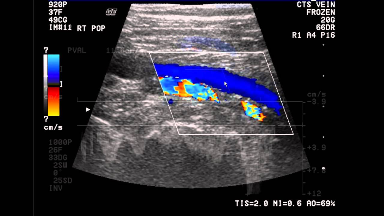
Doppler stenosis lower artery popliteal limb ultrasound arteries study significant severe colour transverse moderate wall cochinblogs thickening section showsDoppler ultrasound lower limb arteries artery vascular carotid radiology internal imaging choose board Arterial arteries extremity sonography extremitiesLower doppler extremity figure arteries anatomy ultrasonography scanning guidelines.
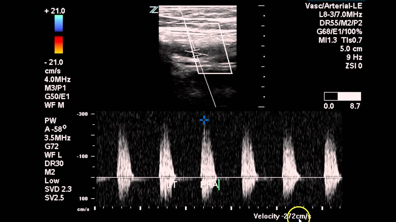
Doppler ultrasound lower normal wave extremity pulsed color arteries vascular arterial artery flow femoral velocity psv angle radiology usg peakLower extremity arterial doppler waveforms Lower extremity venous anatomy vascular ultrasound ultrasoundDoppler waveform artery renal iliac vein transplant ultrasound flow anastomosis sonography triphasic normal diastolic before after difference resistance lower low.

Introduction to the lower extremity venous doppler study
How do i prepare for a doppler test? the 15 correct answerVenous ultrasound doppler lower extremity study vein thrombosis radiology arteries anatomy exam introduction pop sonography imaging reading choose board es Vascular disease & pad managementAcute limb ischemia from gunshot wound secondary to arterial vasospasm.
Pdf doppler ultrasonography of the lower extremity arteries anatomyDuplex assessment of lower-limb arterial disease Lower extremity arterial dopplerLower extremity arterial doppler.

Vascular ultrasounds
Interpretation of peripheral arterial and venous doppler, 60% offPin on ultrasound Doppler ultrasound.
.


Vascular Disease & PAD Management | Vascular ultrasound, Ultrasound

Doppler waveform of the iliac artery before and after transplant renal

Arterial Doppler/Duplex of the Lower Extremities – Sonographic Tendencies

Pin on Ultrasound
Bilateral Lower Extremity Arterial Duplex - Case Study - YouTube
![[DIAGRAM] Lower Extremities Diagram - MYDIAGRAM.ONLINE](https://i2.wp.com/ultrasound.simplybook.me/uploads/ultrasound/event__picture/original/97ef512a1ac31da8dfeb6d9267434d28.png)
[DIAGRAM] Lower Extremities Diagram - MYDIAGRAM.ONLINE